
Testes or scrotal ultrasound is usually carried out when a patient notices a lump or feels pain in his testicles and/or testes area. This is a non-invasive and painless procedure. Using a transducer lubricated with compatible gel, the Radiologist moves the transducer over the scrotum to obtain the necessary images. Both testes and epididymis are examined to characterize any palpable lumps or to identify inflammation in the case of Epididymo-orchitis.
This is a very simple procedure and it does not require any special preparation on the part of the patient.
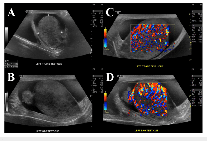
Scrotal lesions can be broadly classified as testicular and extratesticular. The common testicular lesions are torsion, trauma, neoplasms and inflammatory conditions. Extratesticular lesions include lesions of the spermatic cord, epididymis and scrotal wall. In clinical practice, color Doppler imaging of the scrotum is mainly indicated in acute painful scrotal conditions and assessment of varicoceles. Epididymo-orchitis and testicular torsion have similar clinical presentations and color Doppler is useful in accurately differentiating between the two. Testicular viability can also be very well assessed with color Doppler in cases of testicular trauma.
Gray-scale USG is an accurate, fast and useful imaging modality for the imaging of scrotal lesions. Gray-scale USG can distinguish between intratesticular or extratesticular lesions and also aid in the characterization of various lesions. Color Doppler enhances the visualization of varicoceles. Color Doppler USG is the modality of choice to differentiate testicular torsion from inflammatory conditions and can thus help in avoiding unnecessary surgical explorations.