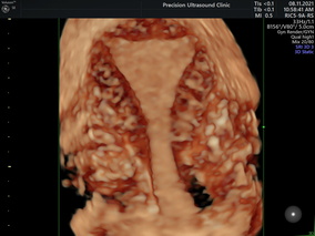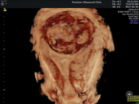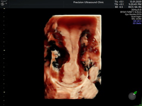
3D Pelvis Ultrasound:– Three-dimensional ultrasound (3D USG) is a fast-evolving imaging technique that holds great potential for use in gynecology. Its sensitivity and specificity are reported to be close to 100 % for diagnosing congenital uterine anomalies. With 3D USG, a coronal view of the uterus can be obtained, clearly outlining the external contour of the uterus and providing accurate information about the shape of the cavity.
Research shows that 3D USG is more sensitive and specific than two-dimensional ultrasound (2D USG) in defining and mapping uterine lesions, such as fibroids, adenomyosis, and intrauterine synechia. In cases of suspected malignancy, 3D USG is mainly used in the initial evaluation of patients. Measuring various indices and mapping vascular architecture with 3D power Doppler have been proposed for evaluating adnexal masses.
In conclusion, clinicians can benefit in adding 3D USG to their routine gynecological workup as it provides fast and accurate results in a relatively cost-effective setting.